


Development of Advanced Dental Implants for Critical Clinical Applications
Investigators: Prof. Emeritus (Dr.) Mahesh Verma, Dr. Sangeeta Talwar, Dr. Farrukh Faraz, Dr. Smiti Bhardwaj, Dr. Preeti Nanda Mishra
Dental implants have become a definitive alternative for tooth replacement. They have epitomized stable long term prosthetic solutions, owing to their conservative benefits, improvised stability and par- natural aesthetic outcomes. However, price of the imported implant systems raises the net – treatment costs and limits their affordability.
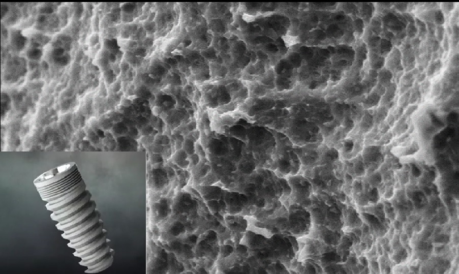
Figuring the need for an affordable implant system, Council of Scientific and Industrial Research (C.S.I.R) under the aegis of New Millennium India Technology Leadership Initiative (NMITLI) initiated a project on development of dental implants, which was led into its Phase I and II trials, under the entrustment of Prof. (Dr.) Mahesh Verma, Maulana Azad Institute of Dental Sciences and Prof. Naresh Bhatnagar, Indian Institute of Technology, Delhi. During this phase, dental implants were exquisitely designed and developed. Their safety and credibility was established through finite element analysis, mechanical testing, bio-compatibility, in - vivo implantation studies and Clinical trials, thus ensuring the highest standards of quality.
The osseointegration ofendosseous implant usually requires 3-6 months. Moreover, osseointegration in challenging situations, such as in immediate implant placement and immediate loading conditions is less predictable and requires bone formation at a faster rate. Considering the current status of global research, it is imperative to obtain indigenous solutions to address the mentioned concerns. For these situations there is a requirement of implants with a paradigm shift from friction closure to form closure and nanotopographic surfaces, which can achieve higher primary stability and fastest possible osseointegration to produce a predictable outcome. Hence, there is need for a next generation of implants, which can be placed in less ideal situations with reduced treatment time. Phase III of the project is concentrating on the enhancements in implant fixture structure, thread design, and surface micro-topography. It will help us develop the technology to address new-age clinical requirements.
Clinical, CBCT Based Radiographic assessment and Biomechanicalanalysis Of Immature Permanent Teeth Treated with Different Regenerative Protocols: Amulti arm Randomized Controlled Trial.
Investigators: Dr.Sudha Yadav, Dr.Ruchika Roongta, Dr. Sangeeta Talwar
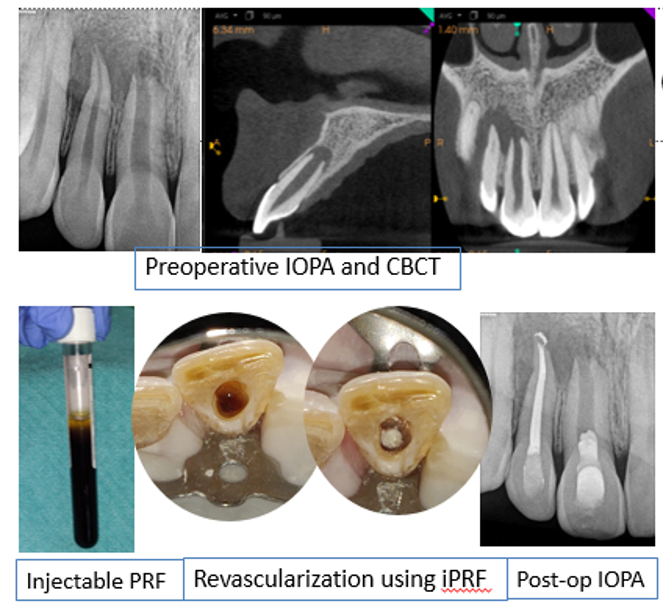
Modern medicine aims to work on two principles viz. prevention and regeneration and endodontics is no exception. In 2007, the Journal of Endodontics published a “call to action” article urging scientists, endodontic professionals, and funding agencies to hasten the development of research in this field of regenerative endodontics. Ever since, this field has received enormous attention from researchers and clinicians. Existing literature shows that both mineral trioxide aggregate (MTA) apical plug (MAP) and regenerative endodontic treatment (RET) have shown acceptable clinical outcomes in immature teeth affected by pulp and the periapical diseases. However, high quality studies directly comparing the outcomes of MAP and RET are scarce. Hence, this Randomized clinical trial aims to compare the clinical and radiographic outcome (using CBCT) of apexification and RET using injectable PRF.
Design and Development of novel instrument retrieval equipment for removal of separated instruments during endodontic therapy
Investigators: Dr. Sangeeta Talwar, Dr.RuchikaRoongtaNawal, Dr. Mahesh Verma, Dr. Amit Malhotra
Endodontic therapy/ Root canal treatment is a common procedure with 80-90% patients reporting in dental OPD requiring it. A possible mishap during this procedure is instrument separation/fracture with reported incidence of 8-10%. Although highly flexible NiTi framework is used to manufacture these instruments, marked complex nature of root canal system induces a stress which sometimes leads to its separation inside the canal. This mishap not just compromises the further course of treatment but is also psychologically traumatic both for the patient and the practioner and may have medicolegal implications. Hence instrument retrieval becomes essential for the success of the treatment. The task of removal requires high degree of skill, specialization and experience. Over decades many systems have been developed with; single frequency ultrasonics yielding fair results so far with a reported success rate of 40-60%. The major drawbacks with the existing techniques are increased treatment time and loss of tooth structure.
Dr. Mridula Goswami
The impact of endodontic microscope on the outcome of root canal treatment of deciduous molar.
Investigators: Dr.Gyanendra Kumar, Dr. Ferah Khanna
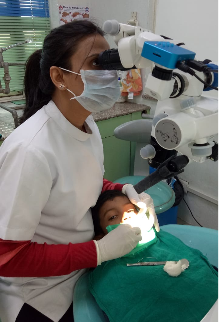
Despite of the recent advances on the preventive front, worldwide prevalence of dental caries is on a rise and seems unlikely to improve in near future causing pulpal involvement in young teeth or their premature loss. Pulp therapy in primary teeth is challenging as it involves thorough cleaning, shaping and debridement of bizarre shaped and tortuous canals with/ without physiologic resorption. Moreover, presence of accessory canal in interradicular region may increase permeability to microbial toxins consequently causing chronic non healing lesions. Thus, it becomes necessary for the dental clinicians to understand the morphology of teeth affected and its adjoining periodontal tissues performing the most appropriate dental treatment in young children. A variety of techniques have been used to study root canal morphology including radiographic examination, root sectioning and staining and clearing techniques. Most of the techniques used to observe the morphology of root canals either alter or destroy the details of the tooth morphology. An important aid for locating root canals is the dental-operating microscope (DOM) which brings minute details into clear view and helps distinguish microstructures that are not visible to the naked eye. It allows the operator to understand the subtleties of pulp chamber anatomy visualize the pulpal floor and locate root canal orifice. Furthermore, the microscope enhances the operator's ability to selectively remove dentine with great precision, locating and negotiating canals, minimizing the procedural error. Thus, it is proposed to assess the impact of Dental Operating Microscope on success of endodontic therapy on primary molars.
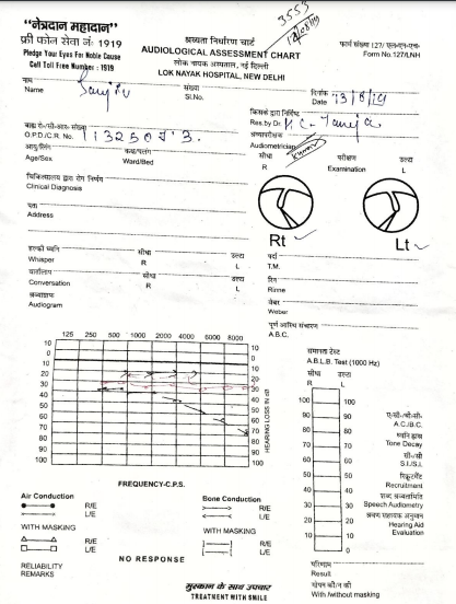
Evaluation of Audiometric Findings, Pre And Post Salivary And Serum Cortisol Levels And Response To Various Treatment Modalities In Myofascial Pain Syndrome
Investigators: Dr. Sunita Gupta, Prof. (Dr.) Mahesh Verma, Dr. Ravi Meher, Dr. Bhawana Mahajan, Dr. Abhishek Kashyap, Dr. Priyanka Verma
Evaluation of Audiometric Findings, Pre And Post Salivary And Serum Cortisol Levels And Response To Various Treatment Modalities In Myofascial Pain Syndrome Investigators: Dr. Sunita Gupta, Prof. (Dr.) Mahesh Verma, Dr. Ravi Meher, Dr. Bhawana Mahajan, Dr. Abhishek Kashyap, Dr. Priyanka Verma
A temporomandibular disorder (TMD) is a term used for Muscle and TMJ disorders. In dental OPD, most TMD patients usually reports with functional disorders of masticatory muscles. In terms of frequency of patients reported with chief complaint of pain, they are only second to odontalgia.Myofascial pain usually arises in muscles at certain hypersensitive areas commonly referred as trigger points. On palpation these trigger points in muscle tissue and/or tendon attachments elicit pain. Little is known about these trigger points. Some suggests these localised zones are created by certain algogenic substances which create hypersensitive areasin theimplicated tissues. Increase in the metabolic demand and/or reduction in blood flow in the tissues may lead to localized rise in temperature at these trigger points.
Local factors such as constant sources of deep pain may also alter function of muscles. Such deep pain may arise from muscles, teeth or joints. Duration of pain seems to be more important than source as muscle response may also be created by idiopathic pain.Normality of muscle function may also be disturbed by systemic factors. Emotional stress is one such systemic factor Muscle function due to the stress may be altered by disturbances in sympathetic activity to the muscle tissues or the gamma efferent system to the muscle spindle.
Increased anxiety and stress leads to additional parafunctional activity in masticatory systemwhich not only increases clenching or bruxing but may increase the generalised muscular tonicity. Besides sress and anxiety, depression is another emotional state associated with TMD.Although, its direct correlation with TMD is not clearly mentioned in literature but chronicity of pain associated with TMD may lead to depression in affected patients. Physiologic stress leads to activation of hypothalamic-pituitary–adrenocortical (HPA) system leading to cortisol secretion from adrenal cortex. Cortisol, commonly known as stress hormone is used extensively for stress evaluation. Higher salivary cortisol levels in the study performed by EmaVrbanovicet et alsuggests that either stress leads to development of TMD, or chronic pain associated with TMD produce excessive stress to the body which further increase cortisol secretion.To the best of our knowledge, this is the first study which is measuring pre and post treatment levels of serum and salivary cortisol.
Very thin boundary lies between TMJ and external auditory meatus and middle ear which can confuse the patient’s ability to locate the pain. Sensation of fullness in the ear or ear stuffiness sensorineural hearing loss,tinnitus, vertigo and altered hearing are the various complaints which have been associated with TMD. There is significant evidence for presence of possible overlapping signs and symptoms between cervical motion impairment or posture differences and TMD which could be explained byanatomical vicinity, neuronal interconnections between trigeminal and cervical.nerves.
Management of TMD start with educating the patient that condition is benign and not aggressive that can threaten one’s life. They should be explained that conservative therapy is usually effective in controlling pain and restoration of function. It is necessary to tell them to protect their temporomandibular joints by restricting the movements within painless limits, eat soft diet, avoid parafunctional habits such as chewing gum and maintain a healthy posture. In treating masticatory muscle disorder one must be aware of emotional stress as one of the etiologic factor. So a holistic approach should be followed which covers patients’ biological, psychological and behavioral aspects. Keeping all these factors in mind combined treatments are being planned to promote relaxation and reduce emotional stress as well as symptoms associated with the condition.
The various treatment modalities which will be used are Occlusal splint therapy, Physical Self-Regulation, TENS and Low Level Laser Therapy. Occlusal splint is a reversible therapy which alters the patient’s occlusal condition temporarily and is effective in reducing muscle pain upto 85%. It provides occlusal stability and decrease the pain and parafunctional habits.Physical self-regulation (PSR) was developed byDrs.Carlson and Bertrand in 1995 to addressing the pain,fatigue, autonomic dysregulation, altered breathing patterns, and improving sleep quality in orofacial pain patients.
TENS is produced its effect by continuous stimulation of cutaneous nerve fibers at a subpainful level. It uses a low-amperage, low-voltage, biphasic current of wide-ranging frequency and is designed primarily for sensory counterstimulation in painful disorders.
Low level laser therapy (LLLT) is most widely used in rehabilitation of nornal function of the tissues. Photochemical reaction takes place when light (photons) enters the cell and reacts with chromophores. It leads to desirable physiologic effects in terms of tissue regeneration, reduction of pain and inflammation.The purpose of this study is a) to study the correlation of aural and cervical findings with myofascial component of temporomandibular disorders exclusively in comparision to previous studies where general term of temporomandibular disorders(TMD) have been used, b) to find out the effectiveness of myofascial pain treatments in patients with aural complaints, c) to find out the degree of stress, anxiety and depression and neck disability amongst these patients.
Evaluation Of Dimensional Changes In The Alveolar Ridge Following Tooth Extraction With Or Without Using Indigenously Prepared Decalcified Freeze Dried Bone Allograft – A Clinical Study
Investigator: Dr. Farrukh Faraz
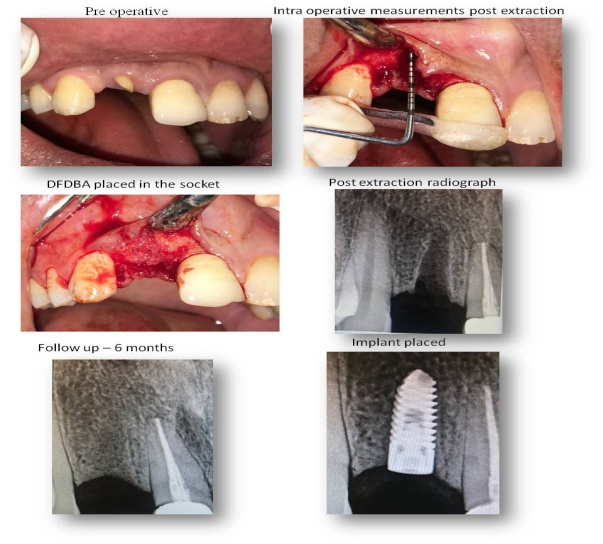
Extraction of a tooth is indicated when it has a poor prognosis and cannot be maintained or restored in an acceptable condition for a long duration. Following extraction, it has been observed, that the alveolar ridge undergoes considerable resorption as a result of a series of biological events triggered by the absence of the tooth. Since the ridge dimension is critical for the placement of implants to replace the lost teeth, several biocompatible materials and autogenous bone grafts have been used to preserve the horizontal and vertical dimensions of the ridge.
The primary aim of this study is to indigenously prepare DFDBA using the international protocols and guidelines and to clinically evaluate the new bone formation following ridge preservation with the prepared grafts.
The 3-year study has been undertaken with the objectives of incorporating the new techniques of graft processing and to provide grafts with a predictable function, free of transmissible diseases.
Evaluation of closure of large alveolar defects by vector controlled transport distractor in cleft lip and palate patients
Investigators- Dr.Tulika Tripathi, Dr. Mahesh Verma, Dr. Navneet Singh
Objective: To assess the treatment outcome of alveolar cleft closure by maxillary alveolar transport distraction using tooth borne distractor in cleft lip and palate patients.
Materials and Methods: A total of 17 subjects were enrolled in thisprospective study, out of which 3 subjects were dropped out. A remaining 14 patients (7 male and 7 female, 10 unilateral and 4 bilateral cleft) were in the age group of 16-25 years. Subjects recruited had large alveolar defect of size greater than width of canine or failure of attempted secondary bone grafting. Presurgical orthodontics involving alignment and expansion of the upper arch was done for visualization of the actual amount of alveolar cleft. The distractor appliance was fabricated on the models of the patients using the Hyrax screw. The arm of hyrax screw was adapted on the cast according to desired distraction vector. A bonded hyrax type of appliance was fabricated with horseshoe shaped acrylic extension over the maxillary dentition with position of hyrax screw at the site of osteotomy cut. Transpalatal Arch was fabricated with 20 gauge wire for reinforcement of anchorage which was embedded into the palatal aspect of the acrylic component of the appliance. Non programmed brackets, and tubes were incorporated on the palatal side of the acrylic portion for ligating 0.18 australian arch wire which provides the guiding channel for transport discs during distraction. The adaptation of distractor was tested clinically before surgery. The appliance may be bifocal or trifocal depending on the amount of defect to be closed
Assessment of salivary Insulin like growth factor-1 and Bone alkaline phosphatase levels with twin block therapy: an observational study.
Investigators: Dr. TulikaTripathi, Dr. Priyank Rai, Dr. Navneet Singh
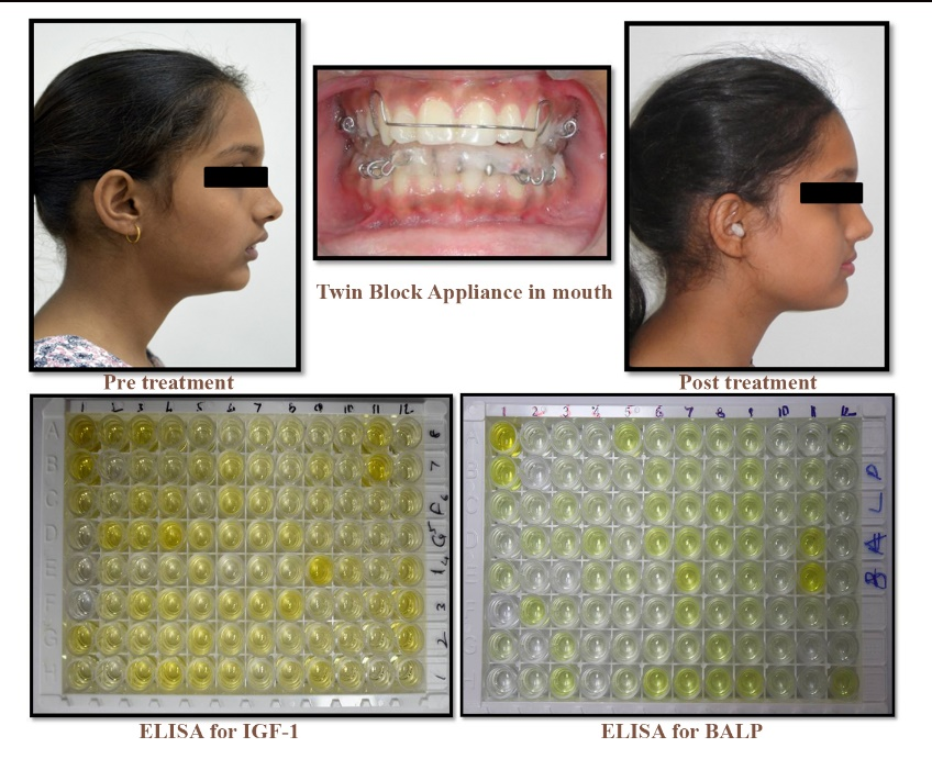
The aim of the study is to assess the changes in levels of salivary IGF-1 and BALP following twin block therapy. This observational study was proposed to be conducted on a minimum 40 subjects having Angle’s Class II division 1 malocclusion with retrognathic mandible in the age group of 10-14 years. Twin block therapy will be started in all the subjects and non compliant subjects will be taken as controls. Unstimulated salivary samples will be collected at various time points for estimation of IGF-1 and BALP by ELISA. The skeletal, dental and soft tissue changes before and after the use of twin block appliance along with condylar changes will be assessed on lateral cephalogram taken at different intervals.
Platelet – Rich Fibrin combined with indigenously prepared Demineralized Freeze Dried Bone Allograft for Treatment of Infrabony Defects in Chronic periodontitis: A clinical Study
Investigators : Dr. Kamal Aggarwal, Dr.Arundeep Kaur Lamba
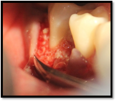
A prospective pre and post interventional comparative study involving patients with a clinical diagnosis of moderate-to-severe chronic periodontitis is being carried out in the department of periodontitis. Inclusion criteria included presence of a 3-walled infrabony defect (IBD) ≥ 3 mm deep in which depth is measured radiographically from the alveolar crest to the base of defect on an intraoral radio-visiographical (RVG) periapical radiograph and the architecture of the 3-walled IBD is confirmed upon surgical exposure of the defect.
Patients are taken up for phase-I therapy which includes thorough supragingival and subgingival scaling and root planning. On re-evaluation after 4-6 weeks of initial therapy full mouth Plaque Index (PI) are recorded and treatment is planned only if the PI is <1.5. Sites for surgical procedure are selected and stents are fabricated for recording of clinical parameters.
Platelet rich fibrin is prepared by taking 10ml of patient’s intravenous blood from median cubital vein in a vaccutainer. Vaccutainer with patients’ blood will be centrifuged at 3000 rpm for 10 minutes by using centrifugation machine.After flap reflection adequate defect debridement and root planning is performed using ultrasonic instrumentation and area-specific curettes. PRF with the bone graft are filled into the IBD.
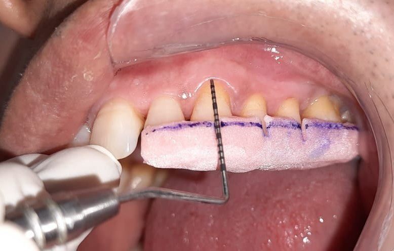
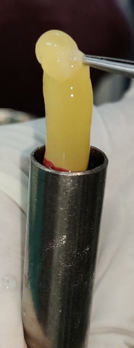
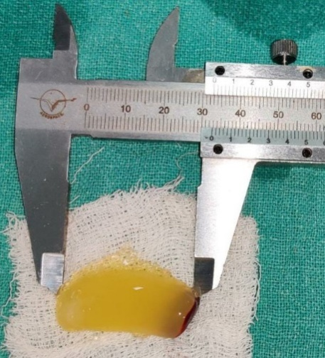
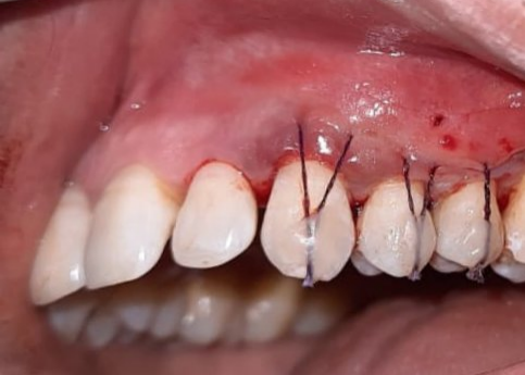
Gum Drop Technique With Titanium Prepared Platelet Rich Fibrin For The Treatment Of Multiple Gingival Recession : A Clinical Study
Investigators: Dr. Archita Datta, Dr. Arundeep Kaur Lamba, Dr.Shruti Tandon
Dental patients of all ages are increasingly concerned about their smile and overall appearance and are particularly unhappy when they have esthetic issues and dental pain associated with exposed roots. Gingival recession is the oral exposure of the root surface due to a displacement of the gingival margin apical to the cemento-enamel junction. Recession seen in healthy patients is mainly due to oral care that is “too much of a good thing” whereas recession seen in periodontally diseased patients is likely due to a chronic inflammatory process that may represent years of “doing too little of a good thing”. Regardless of the frequency & intensity of the oral hygiene, the symptom that brings periodontally healthy and unhealthy patients with recession to the dental clinic is a very unpleasant side effect common to both groups – dentin hypersensitivity.
This study is a prospective pre & post interventional study that involves 40 patients complaining of dentinal hypersensitivity along with at least 2 recession‐type defects affecting adjacent teeth in esthetic areas of the mouth. This technique is minimally invasive and it addresses the goals of root recession coverage with a biological approach to stimulate healing, reduce inflammation during healing, and provide long term stability of the repositioned gingival margin. A less invasive surgical approach has shown higher patient acceptance. Results observed on Patient centered outcomes i.e dentinal hypersensitivity &esthetic score have shown significant difference. The new biological approach of using T-PRF has provided a stimulus to growth factors encouraging new attachment of the gingival tissue to the root surface. This has led to identifying a new economical and readily available alternative to commercially available allografts. The technique has shown success and will work as a foundation step for future research on titanium prepared platelet rich fibrin (T-PRF) as an autologous graft for various therapeutic procedures.
Therapeutic Assessment of Zerumbone/Nanozerumbone in Combination with Curcumin/Nanocurcumin on Oral Cancer Cell Lines.
Investigators: Dr. Sunita Gupta, Dr. Mahesh Verma, Dr. M. Moshahid Alam Rizvi, Dr. Shriya Khera
Oral Squamous cell carcinoma (OSCC) accounts for 1, 45, 000 deaths every year. Regardless of the recent advances, the overall survival rate of the affected patients is still reducing. The recent drugs are now designed to act at a targeted pathway for inhibiting the proliferation and spread of OSCC cells. In the proposed study, we would be assessing the synergistic effect of phytochemicals like Zerumbone, nanozerumbone, curcumin and nanocurcumin on the OSCC cell lines. Zerumbone, a tropical ginger sesquiterpene is preset in subtropical ginger, Zingiberzerumbet Smith that possess anti- inflammatory and anti- growth properties in human cancer cell lines. Another phytochemical, Curcumin (diferuloylmethane), a polyphenol, is the main component of the Indian curry spice turmeric and is known to be a powerful antioxidant with strong anti-inflammatory properties and anti- neoplastic properties. Continuing the role of proteonomics, genomics and the efficacy of these neutraceuticals, the present study is planned to assess the synergistic effect of these phytochemicals and with the invention of their nano components for improving the life and survival rate of patients suffering from Oral Squamous Cell Carcinoma. The following tests were done on the cell line: MTT Assay, Cell proliferation assay, Cell cycle assay, FITCH Annexin assay, wound healing assay, Cell invasion assay. The tests concluded an anti apoptotic, anti-proliferative and anti tumour property. It also showed remedial properties that inhibit cell proliferation, migration and invasion of oral cancer cell lines that acts as a potential candidate in the management of OSCC.
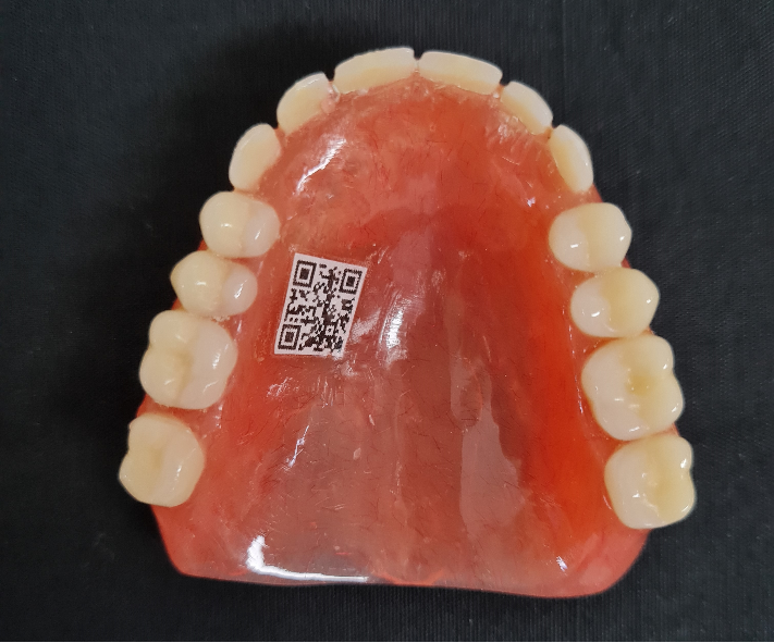
A comparative evaluation of clinical and technical efficacy of denture labelling techniques using Stainless steel band, dynamic Quick Response code and Near Field Communication tags: A Randomized Controlled Trial
Investigators: Dr Modhupa Ghosh, Dr. Rekha Gupta
This project aims at developing an identifiable personal database from a national perspective by using patient’s own dentures as a potential source of information. The purpose is to develop and test a viable denture labelling technique which is simple, cost effective, easily retrievable and based on the modern wireless technologies available. This would allow a patient’s pertinent information to be conveyed to the necessary personnel in time in case of an emergency. This Project is being carried out in the Department of Prosthodontics, MAIDS by Dr Modhupa Ghosh, Senior Research Associate under the guidance of Dr Rekha Gupta, HOD and Professor, Department of Prosthodontics MAIDS and has been funded by CSIR for a period of 3 years.
Comparative Evaluation of Oral Health Status and Treatment Needs of children with Autism Spectrum Disorder (ASD)- A Cross-Sectional Study
Investigators: Dr. Vashi, Dr. Mridula Goswami, Dr. Monica Juneja, Dr. Gyanendra Kumar
Oral health care is essential yet challenging in children with Autism Spectrum Disorder (ASD) due to their impaired emotional and behavioral stability, lack of communication skills and inability to perform daily home hygiene routine properly. The present study was planned with the aim to assess the Oral Health status and Treatment Needs of children with ASD in comparison with children without any systemic disease. A total of 160 children, in the age group of 5-14 years, divided equally into two groups i.e. Group I (children with ASD) and Group II (children without any systemic disease), were assessed for Dental caries, Oral Hygiene Status and Treatment Needs. Behavior of children in each group, during oral examination, was also assessed and recorded. The mean age of participating children was 7.96±2.43 years with a male predominance. Children with ASD displayed more negative behavior with 15% showing definitely negative behavior, 21.2% negative behavior on Frankl’s Behavior Rating scale.Routine oral hygiene care and diet modifications can significantly improve the Oral Health Status in children with ASD.
Efficacy of chitosan-bioglass complex versus sodium Fluoride varnish in remineralization of artificial incipient enamel caries: an in-vitro study
Investigators: Dr. Sonal Bhatia, Dr. Vikrant Mohanty, Dr. Aswini Y Balappanavar
fluoride agents is that they do not contain calcium and phosphate ions and rely on external source, such as saliva, for the remineralization of tooth structures. Bioglass has been show to achieve remineralization of incipient enamel caries. Chitosan along with Bioglass can show better delivery of bioglass and hence we can achieve more targeted remineralization. Thirty-six artificial enamel white spot lesions were created by acidic gel model and subjected to 28d pH-cycling model. Samples were assigned to 2 groups: (1) chitosan-bioglass slurry (CBS) and (2) Fluoride Varnish (FV). Mineral content changes, surface microhardness and ultrastructure were evaluated by EDX, Vickers microhardnessand SEM, respectively. Chitosan-bioglass complexing was found to exhibit greater mineral regain and recovery of surface microhardness compared to "standard" Fluoride varnish after pH-cycling remineralization for 28 days (p < 0.05). Specifically, dense precipitations with Ca/P ratios similar to that in pure hydroxyapatite (HA) were observed which filled the porosities, leaving no prismatic enamel structure exposed. Chitosan-bioglass complex is positive in promoting mineral deposition. Clinically significant chitosan-bioglass formulations may provide analternative clinical strategy in remineralizing early enamel carious lesions as well as desensitization.
Compliance assessment of COTPA section 4, 5 and 6 among High Education Institution in Delhi: A cross sectional study
Investigators: Dr. Radhika Jain, Dr. Vikrant Mohanty, Dr. Aswini Y Balappanavar
Youth are increasingly been involved in tobacco use and HEIs are seen to be a transition place where they are maximally indulged in such adverse health behaviours. Considering this, Government of India enacted Cigarette and Other Tobacco Products Act (COTPA) in 2003 with its key sections 4,5 and 6. However, enforcement of the law is still a matter of concern. Hence, the present study was designed to assess the compliance of COTPA Section 4, 5 and 6 among HEIs in Delhi. A cross-sectional survey was conducted among HEIs affiliated by Guru Gobind Singh Indraprastha University (GGSIPU) in Delhi. Multistage random sampling was used to select 60 HEIs and 500 students studying under these institutions from 5 zones. Compliance assessment and the Knowledge, Attitude and Practice of students regarding tobacco control laws. Overall compliance of HEIs was found to be to be 45.8% with compliance rate for section 4, 5, 6 and 6b were 62.7%, 42.5%, 19.2% and 47.3%. Total 150 POS were found within 100 yards of selected HEIs. Overall good awareness (56%), positive attitude (76%), and poor practice (89%) was reported among study participants regarding tobacco control laws. The study highlighted good compliance towards COTPA section 4 whereas poor compliance for section 5, 6a and 6b among HEIs. Thus, a concerted effort has to be made to increase the compliance and awareness of the act amongst the youth.